| enginuity |
|
home | contents | previous | next |
Here's looking at you, kid
Up to now it has required something of an act of faith to see the image of an unborn foetus in the fuzzy speckled image produced after an ultrasound scan of a pregnant mother. Using the technology developed by Drs Richard Prager and Andrew Gee, this could all change, as shown in the accompanying illustration.
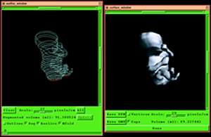
The 3D ultrasound image on the right is produced by reconstructing the surface from a series of line drawings around segments of a foetus as shown on the left, produced using a 2D probe. These techniques can also be used for the volume measurement of internal organs. Such measurements are useful for the monitoring of, say, cancerous growth, or for the calculation of drug dosages.
For more stunning images and an explanation of the techniques that are used, please visit our web site.
| number 9, July 2000 | home | contents | previous | next |