![]()

|
|
||||||||
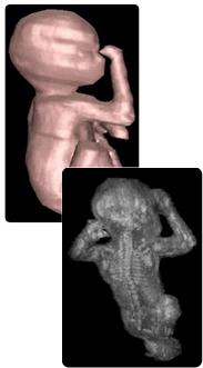
Here's looking at you, kid...
Three Dimension Ultrasound Imaging
|
Ultrasonic scanning is now a standard technique used by doctors to produce images of the inside of the body, the scanning of pregnant women to check on the health of their foetus being the most common application. This technology has been taken a step forward by Drs Richard Prager and Andrew Gee so that three dimensional images can now be produced from a series of two dimensional scans. Not only are the baby pictures much clearer, but this also means the technique can be used for other applications where the volume of an organ may need to be measured to calculate drug dosages or to monitor the progression of a disease such as cancer. For more information on how 3D ultrasound imaging works, click on the button below. |
Animations of foetal scans: |
![]()
![]()