![]()

|
|
||||||||
Three-Dimension Ultrasound Imaging
Anyone who has had a baby has probably seen an ultrasound image. Prospective mothers often come away from the clinic clutching a fuzzy grey picture showing some part of their foetus. In this article we explain how ultrasound imaging works and describe our work on extending it into three dimensions. Ultrasound images are simply a map of the reflected echoes when a pulse of high frequency sound is transmitted into the body - Figure 1 shows a typical example*. White spots in the image show where the sound has been reflected strongly (by bone, for example), whereas darker patches indicate weaker reflections. Echoes which come back first relate to material close to the skin, showing up as bright patches near the top of Figure 1. Later echoes come from deeper in the body and are used to shade more distant parts of the image.
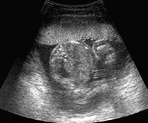
Figure 1: A typical ultrasound image. The surface of the ultrasound probe is at the top of the image and the image extends about 14cm into the body. It shows a slice through the abdomen of a 22 week foetus in utero.
* The authors wish to thank Lois Galletly for her cooperation in the creation of the figures.
![]()
![]()