Ultrasound scanners of the type now routinely used in medical applications first emerged in the 1980s. These types of ultrasound machines are comparatively cheap, costing between £10,000 and £100,000 and they are considered safe, even for scanning the foetus in utero. They are quick and efficient to use, and not unpleasant for the person being scanned. Their main drawback is the speckled nature of the image, and the fact that only a two dimensional slice is produced at any given time. This two dimensional limitation makes it difficult to measure the volumes of structures in the body or to visualise complex anatomy.
 Cambridge
University Engineering Department is the home of the Stradx system
for the acquisition and visualisation of freehand 3D ultrasound. Stradx
has been developed over the last ten years by Drs Prager
, Gee and Treece
in collaboration with Drs Berman and Cash in the University Department
of Radiology at Addenbrooke's Hospital.
Cambridge
University Engineering Department is the home of the Stradx system
for the acquisition and visualisation of freehand 3D ultrasound. Stradx
has been developed over the last ten years by Drs Prager
, Gee and Treece
in collaboration with Drs Berman and Cash in the University Department
of Radiology at Addenbrooke's Hospital.
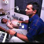
In the Stradx system, a conventional 2D ultrasound machine is used with a position sensor to record the trajectory of the ultrasound probe as the clinician scans. The position information is used to locate each of the 2D ultrasound scan slices in 3D space. Visualisation and volume measurement tools then work on this data. The system was first made available free on the web in 1997 and is now used widely, including groups in Canada, Finland, Germany, France, Spain, Portugal, Hong Kong and the Netherlands. The key features that have led to its leading position in the field are:
In our research, the Stradx system is being principally used in the following applications:
 Talipes
Equinovarus: Clubfoot. This is set of anomalies
in babies' feet involving incorrect tendon lengths and mis-alignments
of the tarsal bone. The failure rate of operative treatment is high and is
confounded by difficulties in imaging tarsal structures which are not yet
ossified and therefore impossible to image by conventional radiology. Freehand
3D ultrasound can be used to image the unossified tarsal bones of the neonatal
midfoot. The figure shows a surface-rendered image, derived from freehand
3D ultrasound, showing the unossified bones in the foot of baby with talipes
equinovarus. Three dimensional animations based on this data together with
geometrical measurements give clinicians a unique insight into the precise
misalignments that are present in each case. This work is almost complete
and two papers have been submitted to Radiology describing it.
Talipes
Equinovarus: Clubfoot. This is set of anomalies
in babies' feet involving incorrect tendon lengths and mis-alignments
of the tarsal bone. The failure rate of operative treatment is high and is
confounded by difficulties in imaging tarsal structures which are not yet
ossified and therefore impossible to image by conventional radiology. Freehand
3D ultrasound can be used to image the unossified tarsal bones of the neonatal
midfoot. The figure shows a surface-rendered image, derived from freehand
3D ultrasound, showing the unossified bones in the foot of baby with talipes
equinovarus. Three dimensional animations based on this data together with
geometrical measurements give clinicians a unique insight into the precise
misalignments that are present in each case. This work is almost complete
and two papers have been submitted to Radiology describing it.
Performance of needle-less injectors. The high resolution facilities of Stradx enable us to reconstruct the distribution of the drug after it has been injected into the body. Results indicate that the high resolution 3D ultrasound system provides significantly better information than magnetic resonance imaging in this application. Our system was used extensively by Weston Medical Ltd to facilitate their design process and the scientific results were published in the British Journal of Radiology.
Spatial mapping of the brachial plexus. Accurate knowledge of the anatomy of the brachial plexus is vital to the successful administration of selective anaesthesia in this area. Using high resolution 3D ultrasound we have demonstrated hitherto unsuspected variations in anatomy which explain past problems administering anaesthetic in this area, and pave the way for safer procedures to be used in the future. Our research registrar, Dr Charlotte Cash, recently won a best paper prize for her presentation on this work at a meeting organised by the European Society of Regional Anaesthesia.
Freehand 3D ultrasound and surface contour mapping to guide breast surgery. The 3D scan information is combined with laser scan information of the surface to produce a 3D interactive visualisation of the location of the tumour under the skin to aid surgery planning. Ethical approval was granted in December 2003 and preliminary trials are underway involving volunteer patients.
Freehand 3D ultrasound guided breast radiotherapy. Freehand 3D ultrasound provides useful information about the location of tissue that was close to the tumour before it was excised from the breast. It can be used to improve the accuracy of the radiotherapy-planning CT scan. If the target of the radiotherapy can be located more accurately in this way, then a reduced overall radiation dose can be used, leading to better cosmetic results.
Freehand 3D ultrasound to measure the volumes of breast tumours as they respond to chemotherapy. Large tumours in the breast are sometimes treated with drugs to reduce their size before surgery is attempted. It is important to measure the way the size of the tumour changes in response to the drug treatment. This is difficult to do as more than one sweep of the ultrasound probe is often required to visualise the whole tumour. The 3D freehand system can overcome this problem and a trial of the system in this application is due to start in June 2004.
For more details see the research group home page.