![]()

|
|
||||||||
Three Dimension Ultrasound Imaging
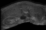
If a scan is performed so that all the slices are approximately in the same plane, the position information can be used to patch them together to create a composite "panorama'' image. This can be done 'live', with the image built up on the screen as the scan is performed (see Figure 5).
|
Figure 5: An extended field of view image generated by joining up a large number of individual 2D scan images. Click here for a larger image. |
It is interesting to draw parallels between today's 3D ultrasound research and the pioneering days of medical ultrasound. The first ultrasound scan was produced by Wild & Reid in 1953. In 1964, Smiths Industries on Clydeside produced the first commercial 2D scanner, called the Diasonograph, incorporating mechanisms from shipbuilding and a multitude of valves. By the 1970s, improvements in transducer design resulted in better images, and the 2D ultrasound machine, as we now know it, emerged in the 1980s.
![]()
![]()