![]()

|
|
||||||||
Three Dimension Ultrasound Imaging
Our research has focused on developing a more accurate approach to volume measurement, based on three dimensional ultrasound. A commercial 2D ultrasound machine can give us images representing a number of slices through the structure of interest. To find the volume, we need to know the relative position and orientation of these slices in three dimensional space. This information can be obtained by attaching a position sensing device to the ultrasound scanner probe. We use a magnetic tracking system. These trackers were originally built for the animation and virtual reality industries. They can be attached to clothing and used to analyse the motion of people's limbs as they walk and run about. They consist of a fixed transmitter with three orthogonal coils of wire (about a 2 inch cube), and a receiver (about half an inch in diameter), similarly containing three smaller coils of wire. The transmitter generates harmless magnetic fields, that are picked up by the receiver and used by the instrument to calculate their relative position and orientation. We mount the transmitter in a fixed location close to the patient, and attach the receiver rigidly to the probe of the ultrasound machine (see Figure 2).
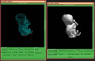
Using the magnetic tracker, we are able to obtain a sequence of slices through the structure of interest at known positions and orientations. Calculating the volume involves drawing a line around the structure in each of the slices and then using an algorithm called "shape-based interpolation" to join the lines in 3D space (see Figure 3). The closed shape thus obtained gives a volume estimate, accurate to within 5%.
|
Click here for a larger image. |
Figure 3: the left image shows the segmented boundaries outlining the body of a foetus in 3D space. The right image shows a surface reconstructed from these boundaries using shape-based interpolation. |
![]()
![]()