![]()

|
|
||||||||
![]()
Three Dimension Ultrasound Imaging
You can probably imagine that the most time consuming part of this process is drawing round the structure to segment it out of each of the image slices. Many groups, including our own, have tried to develop computer algorithms to automate this task, but because of the speckled, noisy nature of the images, none have been successful. Algorithms can be designed to work in certain specific cases, but in general, the only way clinicians can have confidence in the volume estimate is if they are involved in outlining the structure on which it is based. Rather than trying to automate the segmentation, we have concentrated on making the shape-based interpolation algorithm robust so that it works correctly on a relatively small number of manually outlined boundaries. For a typical organ or internal structure, 10 to 20 outlines are enough for an accurate volume estimate. This takes a clinician about 3-7 minutes to segment.
|
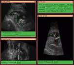
Figure 4: A non-planar reslice, showing a foetus' leg flattened out to be displayed on one plane. Click here for a larger image. |
|
The arrangement of a conventional 2D ultrasound machine with a position sensor attached to the probe has other uses. If the images, and matching positions, are recorded fast so that a dense block of data is obtained, it is possible to calculate slices that cannot be obtained by direct scanning, for example, slices entirely beneath the skin. Non-planar slices can also be computed to "flatten out'' curved parts of the body and make certain aspects of the structure easier to understand (see Figure 4).
![]()
![]()