![]()

|
|
||||||||
![]()
Three Dimension Ultrasound Imaging
There are other ways of producing images of the inside of the body, but they are generally either more expensive than ultrasound, or they involve exposure to X-rays or other radiation. Ultrasound machines are comparatively cheap, costing between £10,000 and £100,000. They are considered safe, even for scanning the foetus in utero. They are quick and efficient to use, and not unpleasant for the person being scanned. Their main drawback is the speckled nature of the image, and the fact that only a two dimensional slice is produced at any given time.
The main clinical consequence of this two dimensional limitation is that it makes it difficult to measure the volumes of structures in the body. This is a common requirement as volumes are used to assess the progression of disease or its response to treatment. They are also useful for calculating drug dosage. Current practice for volume measurement generally involves the use of heuristic formulae. A number of key dimensions are measured and used to provide a volume estimate by combining them with scaling factors learned from previous experience. These techniques can be incorrect by as much as 50%.
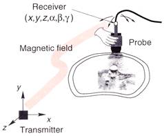
Figure 2: The general configuration of a freehand 3D ultrasound system, showing the probe of the 2D ultrasound machine and the magnetic position sensor.
![]()
![]()