| (a) | 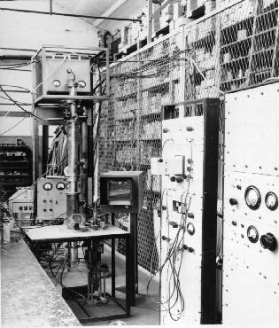 |
(b) | 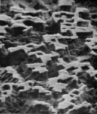 |
His first research student, D McMullan, commenced in 1948, and a working instrument was produced by 1951 (Fig 2(a)). This incorporated three features which differentiated it from its predecessors: electrons scattered from the specimen surface were detected by means of an electron multiplier with beryllium-copper dynodes; the specimen was placed at a large angle to the electron beam; and a high primary beam energy was used to reduce the effects of surface contamination on secondary emission. In addition, images were viewed directly on a slow-scan radar-type CRT display, and recorded on film using a short persistence high resolution CRT. This instrument from the start produced the striking `three-dimensional' images characteristic of the modern-day SEM (Fig. 2(b)).
| (a) | |
(b) | |
| Fig. 2. (a) McMullan’s original microscope, SEM1. (b) Early micrograph (etched aluminium) obtained with SEM1. Horizontal field width: 37 µm. |
The results obtained with SEM1 convinced Oatley, even at this early stage, that the SEM would turn out to be an important scientific laboratory tool; however, this was far from being the consensus among microscopists who, with notable exceptions, believed that this new instrument could never compete with the well-established electron microscopical techniques of the time; in particular, with the replica technique which offered much superior resolution. The new instrument was met with indifference and even, in some quarters, ridicule. Undeterred, Oatley decided to continue with the project. His main argument for doing so was that for a very large range of specimens, high resolution was not important and moderate magnifications sufficed to provide the all-important information. The SEM offered ease of specimen preparation, large depth of field, readily interpreted images, and great flexibility in the size and type of specimen that could be examined, including the observation of dynamically changing specimen properties. All of these advantages were appreciated by Oatley on the basis of the first results obtained with SEM1 - a full decade before microscopists in general were forced to the same conclusion. Two more research students were taken on: K C A Smith in 1952, who continued with the development of SEM1, and O C Wells a year later, who embarked on the construction of a new microscope, SEM2. L R Peters, a gifted technician, was assigned to the project.
Although the electron multiplier appeared to offer an elegant solution to the problem of detection in the SEM, its implementation proved to be technically extremely difficult. Detection of the low-energy secondary electron component required the input of the multiplier to be at a positive potential of several hundred volts, which in turn necessitated the output to stand at about 6 kV above ground. Because capacitors operating at high voltage generate significant noise, capacitive coupling of the signal to ground level at this stage was not possible. Consequently, a head amplifier floating at 6 kV, with all its attendant power-supply, screening and decoupling problems, had to be used to boost the signal to a level at which a capacitor noise became insignificant. Capacitive coupling also necessitated the use of beam blanking in the column and d.c. restoration circuits to maintain black level. All of these difficulties were eliminated at a stroke with the introduction in 1956 of the ‘Everhart-Thornley’ detector. With this detector the output of the multiplier is at ground potential, and d.c. coupling can be used throughout the whole signal chain.
How this detector evolved as the research in Oatley’s Group
progressed is illustrated in Fig. 3. It was while Smith was experimenting with an
environmental cell that Oatley first suggested the use of a plastic organic scintillator
coupled to a photomultiplier to detect the electrons transmitted through the cell. In
Oatley’s arrangement (Fig 3(a)) the rod-shaped scintillator forms one wall of the
cell, and acts as a light-pipe to convey scintillations to the photocathode of the
multiplier.
|
|
Fig. 3. Evolution of the Everhart-Thornley detector. (a) Oatley’s arrangement for detecting electrons transmitted through environmental cell. (b) Detection of BSE component. (c) Everhart’s SE detector. (d) Electron trajectories determined by Thornley. |
This detector was used subsequently by both Wells and Smith to produce backscattered electron images of solid samples (Fig 3(b)). Everhart then took over SEM1 and extended the technique to the detection of the low-energy secondaries (Fig 3(c)). A little later Thornley conducted a detailed study of Everhart’s detector using an electron trajectory tracer (Fig. 3(d)) - another research project initiated by Oatley (Sander 1951). The wide-ranging nature of the programme of research and development that Oatley organised to demonstrate the power and flexibility of the SEM is perhaps best illustrated with reference to the work of his students who participated in the programme. A brief summary of their work is given below. The period spans from the late 1940s to the early 1960s, when the weight of evidence in favour of commercial production of the SEM finally became overwhelming. A full account of the work undertaken in this period, together with a complete list of references to published work, is given in Oatley et. al. 1985.
D McMullan: 1948. Constructed SEM1 and produced the first micrographs showing the ‘three-dimensional’ topographic image formation characteristic of modern-day instruments. Theoretical analysis of probe formation; measurements of distribution of backscattered electrons with angle; considered mechanisms of BSE contrast formation.
Dissertation: "Investigations relating to the Design of Electron Microscopes"
1952.
K C A Smith: 1952. Made improvements to SEM1, including efficient detection of the low-energy secondary electron (SE) component; considered SE contrast formation; extended theory of probe formation; investigated a wide range of applications; dynamic experiments, including chemical reactions at elevated temperatures.
Dissertation: "The Scanning Electron Microscope and its Fields of
Application" 1956.
O C Wells: 1953. Constructed SEM2 and applied it to study of fibres. Explored new types of detector; used scintillator/photomultiplier detector in many novel configurations; established theory of stereomicroscopy in the SEM; investigated ways of examining non-conducting specimens, including the use of positive ion bombardment; investigated atomic number contrast.
Dissertation: "The construction of a Scanning Electron Microscope and its
application to the study of fibres" 1957.
T E Everhart: 1955. Continued improvements to SEM1; devised new detector (‘Everhart-Thornley’ detector). Studied contrast mechanisms in detail, including potential contrast; developed a new theory of reflection of electrons from solids.
Dissertation: "Contrast formation in the Scanning Electron Microscope" 1958.
P J Spreadbury: 1956. Constructed a simple SEM using a cathode-ray oscillograph as the display unit; made careful measurements of the performance of the electron gun. Constructed numerous pieces of electronic equipment which were used in other SEMs and projects in the laboratory.
Dissertation: "Investigations relating to the design of a Simple Scanning Electron
Microscope" 1958.
R F M Thornley 1957. Made improvements to SEM2. Conducted detailed study of Everhart detector. Low-voltage operation for examination of non-conducting specimens; studies of frozen biological specimens.
Dissertation: "New applications of the Scanning Electron Microscope" 1960.
A D G Stewart 1958. Completed construction of a new microscope begun by Oatley (SEM4). Addition of ion gun allowed the direct observation of specimens while undergoing sputtering. Later moved to Cambridge Instrument Co. to work on ‘Stereoscan’ project (see section 5).
Dissertation: not submitted.
H Ahmed 1959 (supervisor A H W Beck). Used SEM2 to investigate activation processes of dispenser cathodes. Direct observation of emitting cathodes at temperatures exceeding 1300 K.
Dissertation: "Studies on high-current-density Thermionic Cathodes" 1962.
R F W Pease 1960. Designed a new microscope (SEM5) - first SEM to achieve a resolution of 10 nm. Several of these instruments were made in the Engineering Dept. and used in other Groups. Also supplied to F P Bowden’s Group in the Cavendish Laboratory (see section 5).
Dissertation: "High resolution Scanning Electron Microscope" 1963.
A N Broers 1961. Made improvements to SEM4, including addition of magnetic lens. Added mass-filter to ion beam system to obtain pure ion beam species. Found that surface contaminants affected rates of sputtering and could thus be used to mask selected areas of the specimen surface. Used this effect to lay down patterns of gold wires and other structures - one of the earliest successful attempts at electron beam microfabrication and micromachining.
Dissertation: "Selective ion beam etching in the Scanning Electron
Microscope" 1965.
Oatley was appointed to the Chair of Electrical Engineering and Head of the Electrical Division in the Department in 1960, and thereafter his direct involvement with the research programme declined; supervision of Pease and Broers was handed over to W C Nixon, who had joined the Department from V E Cosslett’s Group in the Cavendish in 1959. On his retirement in 1971, however, Oatley again took up the research and continued to make contributions to the field well into his 80s (Oatley 1975, 1981, 1983, 1985). His book on the SEM was published in 1972.
5. COMMERCIAL DEVELOPMENT: THE STEREOSCAN
Up to the mid-1950s there was little interest in the work of
Oatley’s Group, but a turning point in attitudes towards scanning electron microscopy
came when D Atack, a member of the Pulp and Paper Research Institute of Canada (PPRIC),
and J H L McAuslan, with Imperial Chemical Industries, learned of the work on the SEM.
Both were on sabbatical leave at the time in F P Bowden's Group in the Cavendish, and they
decided to explore the potential of the instrument for their work. Using SEM1 Atack
examined a range of pulp and paper specimens, while McAuslan studied the thermal
decomposition of silver azide crystals. This work generated strong interest at the PPRIC,
and it was subsequently arranged that the Institute would finance the construction of a
new SEM in the Engineering Department. Smith undertook the task of designing this new
instrument - designated SEM3 - as a post-doctoral research project in 1956.